triceps brachii posterior view
Contents 1 Structure 11 Innervation. The tunica is a strong tendon-like tissue that surrounds all three corpus chambersand the smooth muscle within the chambers.
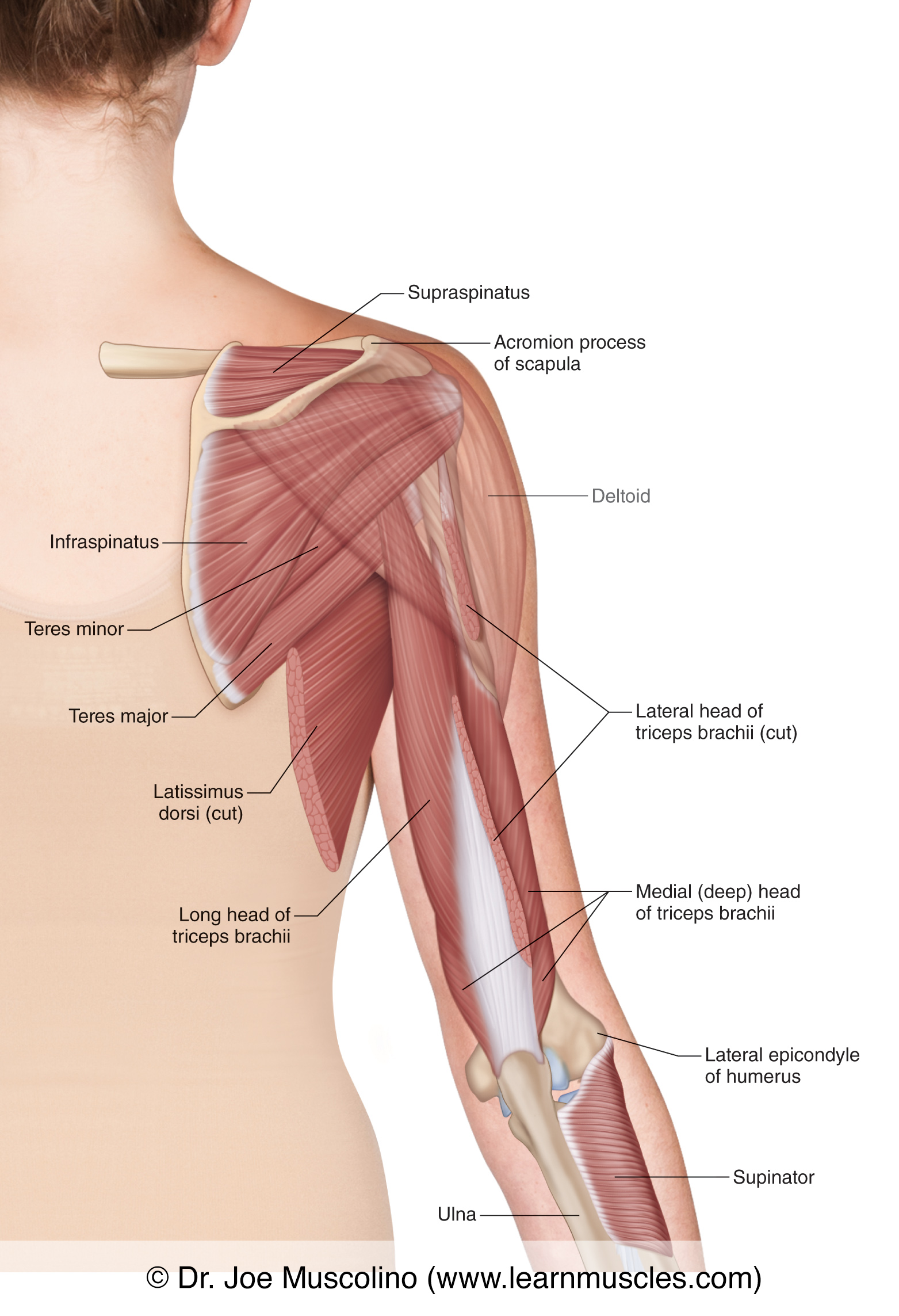
Muscles Of The Posterior Arm Deep View Learn Muscles
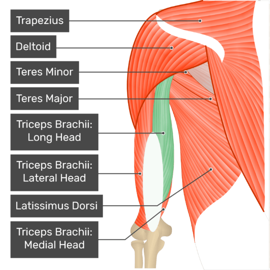
Correctly label the following muscles of the posterior view.
. A long head a lateral head and a medial head. Start studying triceps brachii. The Smooth Muscle the Tunica.
Duquin T Bisson L. 1697704906 Triceps Brachii Muscles Isolated in Posterior View Anatomy 3D Rendering on White Background Formats 8000 8000 pixels 267 267 in DPI 300 JPG 1000 1000 pixels 33 33 in DPI 300 JPG 500 500 pixels 17 17 in DPI 300 JPG Show more Contributor Hank Grebe. Posterior view of the triceps brachii and related nerves.
It consists of 3 parts. The superficial triceps insertion is split lon-gitudinally dark arrows and partially detached from the inser-tion on the olecranon. Ga225007 Fotosearch Stock Photography and Stock Footage helps you find the perfect photo or footage fast.
Triceps Brachii Gluteus Maximus Gastrocnemius Biceps Femoris Trapezius QuestionLabel the muscles in this posterior view of a muscle model. Lateral and posterior surfaces of proximal 12 of body of humerus and lateral intramuscular septum. We feature 66600000 royalty free photos 337000 stock footage clips digital videos vector clip art images clipart pictures background graphics medical illustrations and maps.
The triceps brachii is a large thick muscleon the dorsal part of the upper arm. Posterior surface of olecranon process of ulna and antebrachial fascia. The triceps brachii crosses the elbow joint.
The triceps or triceps brachii Latin for three-headed muscle of the arm is a large muscle on the back of the upper limb of many vertebrates. Triceps Brachii Gluteus Maximus Gastrocnemius Biceps Femoris Trapezius. Posterior view of the elbow with the humerus above and the ulna below.
4 muscle fibers of the medial head of the triceps. Also seen are the supraspinatus infraspinatus and teres minor of the rotator cuff group and the teres major latissimus dorsi cut and supinator. Duquin T Chavan P Bisson L.
A triceps split approach may allow more visualization and exposure of the posterior joint and therefore lessen the need for tri. The medial lateral and long head. Am J Sports Med.
As the name implies it has three heads. Whilst all three arise from different origins they all come together. In this deep view of the right-side posterior upper arm we see the deltoid ghosted and triceps brachii long head is cut.
Posterior brachial cutaneous nerve branch from radial nerve emerges from around the front of the long head of triceps to become posterior near the intersection with teres major posterior circumflex humeral artery and axillary nerve through quadrangular space lateral to the long head of triceps teres major anterior to the long head of triceps. The main function of the triceps is the extension of the elbow joint. Lets start with the tunica.
The main function of the triceps is the extension of the elbow joint. Learn vocabulary terms and more with flashcards games and other study tools. This picture starts with an outer view on the left and then goes deeper towards the right.
Triceps brachii is situated on the posterior aspect of the arm and arises from three heads - the lateral medial and long head. Also its hard to tell but the tendon on the thumb is highlighted too. A posterior view of the muscles of the upper arm relative to the skeleton.
The triceps brachii and the anconeus. The main function of the triceps is the extension of the elbow joint. Which Method of Rotator Cuff Repair leads to the Highest Rate of Structural Healing.
The triceps brachii often shortened to just triceps is located on the posterior portion of the upper arm. The finding that the distance from the articular tip of the olecranon to the proximal tendon insertion is greater. Click on image for larger view.
It often appears as the shape of a horseshoe on the posterior aspect of the arm. It is composed of three heads tri three cep head. Infraglenoid tubercle of scapula and joint capsule of the shoulder long head oblique ridge in the upper half of the posterior humeral shaft lateral head posterior and medial surfaces of the humeral shaft below the point of insertion of the teres major to just proximal to the trochlea medial head Insertion.
The triceps mechanism consists of 2 components the tri-ceps properthat portion inserting on the olecranonand Figure 2. The triceps brachii is a large thick muscle on the dorsal part of the upper arm. Triceps brachii muscle Musculus triceps brachii Triceps brachii is a three-headed tri - three cep - head muscle of the armIt represents the only constituent of the posterior muscle group of the arm spanning almost the entire length of the humerusThe triceps brachii muscle consists of a long medial and lateral head that originate from their respective.
It is composed of three heads tri three cep head. The tunica governs the size of the erect penis similar to how a bicycle tire limits the expansion of the inner tube inside. Innervation of the Supinator Muscle An Anatomic Study.
View the full answer. In the posterior elbow compartment two muscles cross the posterior surface of the elbow. Posterior view near the olecranon insertion.
It is the muscle principally responsible for extension of the elbow joint straightening of the arm. Duquin T Sperling J. It is positioned between the shoulder and elbow joints and works as an antagonist to the.
The triceps brachii is a large thick muscle on the dorsal part of the upper arm. And yes if you look closely you can read the name of the muscle Appendicular Day 2 ONLINE without repea. Appendicular Day 1 ONLINE without.
Deep Superficie Rhomboid major Sternocleidomastoid Rhomboid major Gastrocnemius cut Adductor magnus Gluteus minimus Semimembranosus Flexor digitorum longus Triceps brachii cut Fibularis longus Rhomboid minor Reset Zoom. A long head a lateral head and a medial head. 3 distal pre-tricipital space.
It often appears as the shape of a horseshoe on the posterior aspect of the arm. The long head originates from the infraglenoid tubercle of the scapula. Stock Illustration - LifeART.
Science Anatomy and Physiology Anatomy and Physiology questions and answers Label the muscles in this posterior view of a muscle model. 5 tendon and muscle fibers of the long head of the triceps strip. It often appears as the shape of a horseshoe on the posterior aspect of the arm.
The triceps brachii is shown.

