Best Way to Use This Resource. Lymphatic Drainage of the Larynx.

Pin On Images For Voice
It consists of a cartilaginous skeleton connected by membranes ligaments and associated muscles that suspend it from.

. Leaf-like cartilage that drops to cover the opening to the larynx during swallowing. Strap muscles anterior jugular vein. Larynx lateral view right side.
11 and 12 The cricoid cartilage lies between the thyroid cartilage and the trachea. Medial view of the right side of the larynx. Label the blood vessels of the female pelvis using the hints provided.
Posteriorly it has a large quadrate-shaped lamina that narrows into the arch anteriorly. 4 rows This is an online quiz called Posterior View of the Larynx. Membrane that spans the space between hyoid bone and the lateral thyroid.
The bone shared by the tongue and the larynx. View the full answer. Of the larynx except the cricothyroid and sensation to the larynx below the vocal cords.
Need to osmose some anatomical knowledge. There is a printable worksheet available for download here so you can take the quiz with pen and paper. The form of the lateral aspects is determined by the larynx cartilages and consist of three parts a superior one that matches the thyroid cartilage an inferior one that matches the cricoid cartilage and a middle.
950 The cartilages of the larynx. 12 Posterior view of the larynx. Larynx anterior view The larynx is a complex hollow structure located in the anterior midline region of the neck.
It is situated between the trachea and the root of the tongue at the upper and. Lymphatic Drainage Lungs Innervation of Bronchial Tree. This specimen comes fresh from the morgue so you can test your knowledge all the way to the exam room.
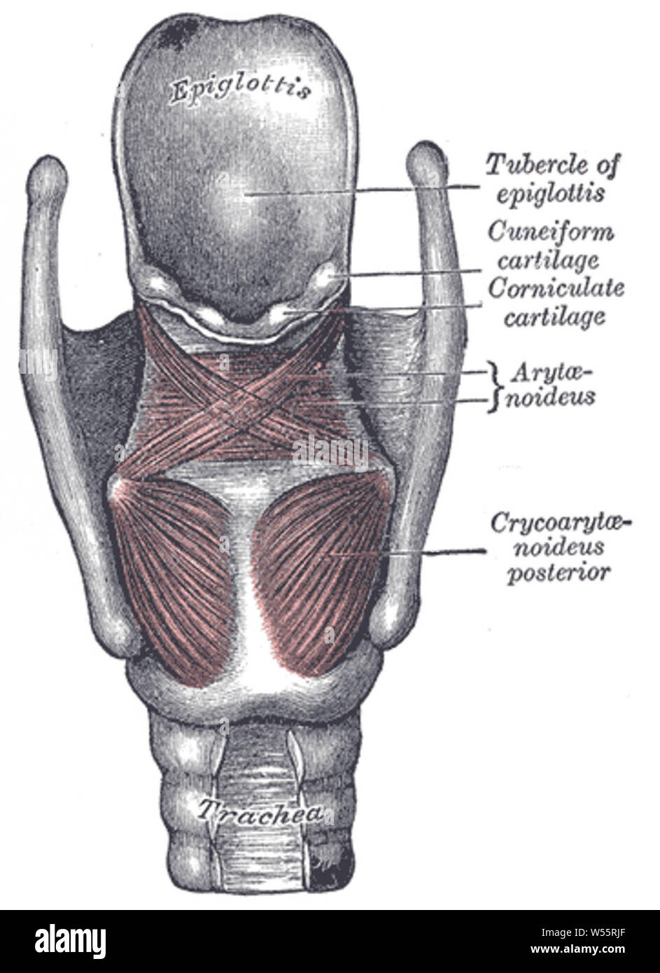
Its anterior part is called the band. View of the larynx at laryngoscopy Base of tongue Glossoepiglottic fold Vallecula Epiglottis Epiglottic tubercle False cord Vocal fold Vestibular fold Aryepiglottic fold Cuneiform cartilage Corniculate cartilage Trachea Figure 3. This high resolution full color PDF Digital Download of the larynxs posterior view is just what the doctor ordered.
As it extends upward to form the posterior border of the larynx it tapers anteriorly to a narrow arch. The cricoid is attached to the top of trachea to the first tracheal ring by the cricotracheal ligament. Label the blood vessels of.
The epiglottis is elastic cartilage shaped like a spoon that is posterior to the root of the tongue. Learn vocabulary terms and more with flashcards games and other study tools. The cricoid cartilage provides attachments for the various muscles cartilages and ligaments involved in opening and closing the airway.
The posterior commissure of the larynx is a name often given to the posterior portion of the glottis. 100 8 ratings The anterior view of the larynx labeled The posterior view of the larynx labeled The epiglottic cartilage. When only air is flowing into the larynx the inlet to the larynx is open wide with the free edge of the epiglottis projecting superiorly and anteriorly.
Terminology The term commissure is a misnomer as the true vocal cords do no. It is anterior to the esophagus and at the level of the third to the sixth cervical vertebrae in its normal position. The larynx or organ of voice is placed at the upper part of the air passage.
Venous Drainage of the Lungs. A portion of the left half of the larynx above the cricoid cartilage and the muscles have been removed. It extends from the upper border of the thyroid cartilage to the greater wing of the hyoid bone.
The posterior part of the internal space of the larynx is part of the anterior wall of the pharynx and has two vertical recesses referred to as the piriform sinus. The interarytenoid muscles are part of this anatomical landmark. Part of the thyroid cartilage has been removed on the right side and the cricothyroid muscle divided.
The thyroid cartilage has been. This is an online quiz called Anterior and Posterior view of Larynx. The lower end of the epiglottis is attached to the deep surface of the thyroid cartilage.
It forms the lower part of the anterior wall of the pharynx and is covered behind by the mucous. Its primary function is to protect the lower aviation by abruptly closing on mechanical stimulation thereby interrupting breathing and preventing the entry of foreign matter into the airway. Superior horn of thyroid cartilage.
There is a printable worksheet. Extends from the inferior surface of the true vocal cords to the inferior aspect of the cricoid cartilage. In contrast to the thyroid cartilage it is a complete ring signet in shape.
Label the structures seen in the anterior and posterior view of the larynx. Various parts of the larynx area closed by connective tissue membranes. The thyrohyoid membrane was seen in the study of the neck and is pierced by the internal laryngeal nerve and superior laryngeal artery.
Forepart of the neck where it presents a considerable projection in the middle line. Posterior view of larynx The larynx is located inside the front appearance of the neck front of the lower portion of the pharynx and above the trachea. Label the posterior view of the larynx based on the hints if provided.
Well look no further. Start studying Cartilages of the Larynx posterior view. Blood Supply to Lungs.
Innervation of the Larynx.

Posterior View Of The Larynx And Vocal Cords Bones Muscles Cartilages Stock Photo Alamy

1 5 Posterior View Of Larynx Showing Aryepiglottic And Oblique Download Scientific Diagram

Posterior View Of Larynx Human Body Vocabulary Medical Knowledge Medical Anatomy
Posterior Larynx Anatomy With Annotations High Res Vector Graphic Getty Images
Muscles Of Larynx Posterior View Stock Photo Alamy

Schematic Of The Human Larynx Framework Based On Gray 6 A Download Scientific Diagram

Anterior View Of Larynx Download Scientific Diagram
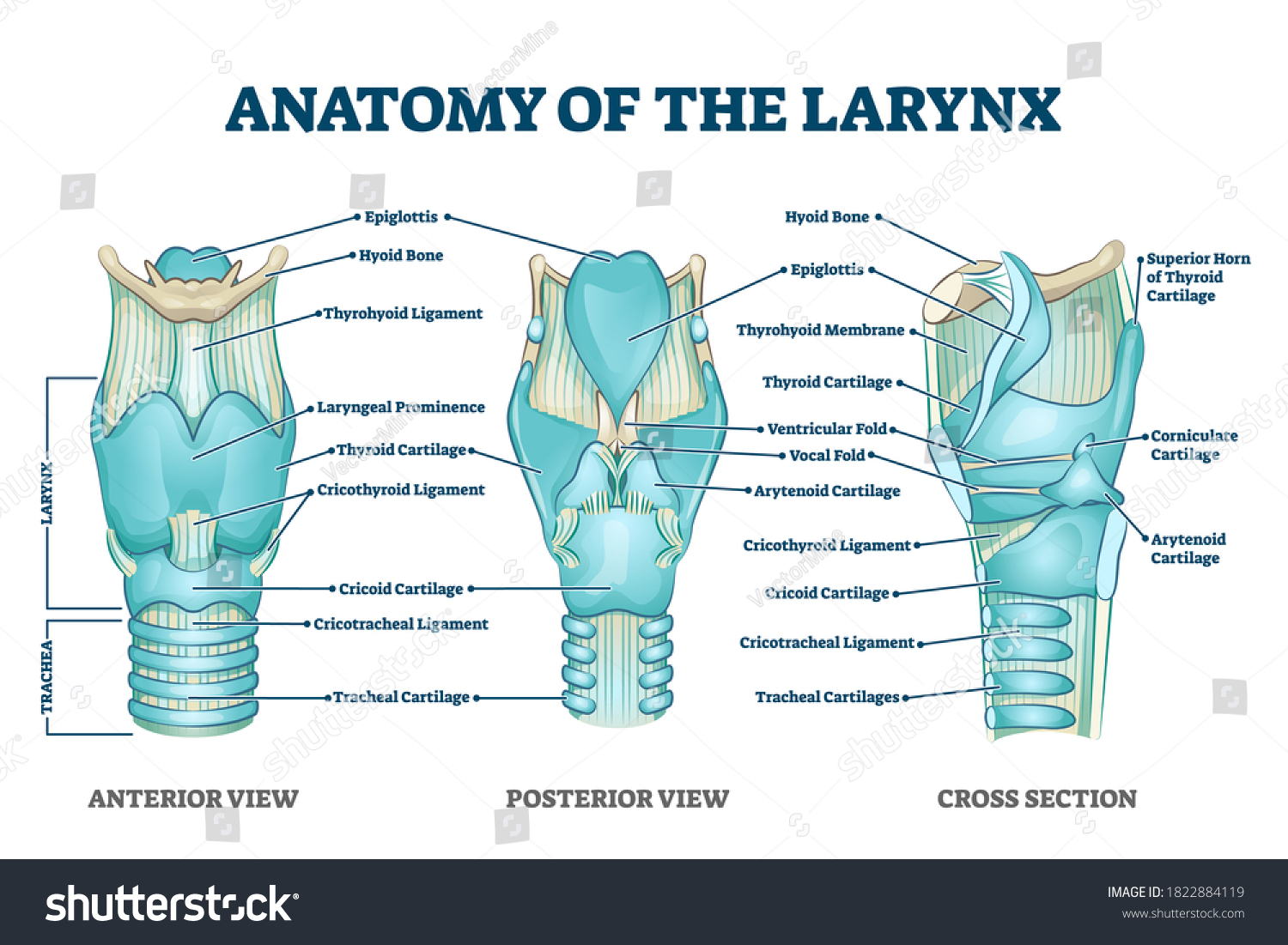
Vektor Stok Larynx Anatomy Labeled Structure Scheme Educational Tanpa Royalti 1822884119
0 comments
Post a Comment